45 labels of the human brain
Human Brain Photos and Premium High Res ... - Getty Images Browse 27,560 human brain stock photos and images available, or search for human brain anatomy or human brain illustration to find more great stock photos and pictures. human brain icons - vector illustration - human brain stock illustrations. abstract brain activity image - human brain stock pictures, royalty-free photos & images. 3D Brain This interactive brain model is powered by the Wellcome Trust and developed by Matt Wimsatt and Jack Simpson; reviewed by John Morrison, Patrick Hof, and Edward Lein. Structure descriptions were written by Levi Gadye and Alexis Wnuk and Jane Roskams .
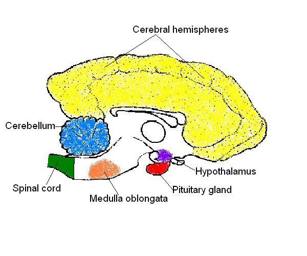
BIO201-Human Brain - Savalli The Human Brain. Major structures on the human brain. Although cranial nerves are shown here by Roman numeral, you should also be able to identify each of these by name and you should know their function. Return to unlabeled photos. This page last updated 16 October 2013 by Udo M. Savalli
Labels of the human brain
Labeled brain anatomy Images, Stock Photos & Vectors Find Labeled brain anatomy stock images in HD and millions of other royalty-free stock photos, illustrations and vectors in the Shutterstock collection. The Human Brain - Visible Body The brain gives us self-awareness and the ability to speak and move in the world. Its four major regions make this possible: The cerebrum, with its cerebral cortex, gives us conscious control of our actions. The diencephalon mediates sensations, manages emotions, and commands whole internal systems. The cerebellum adjusts body movements, speech ... 101 Labeled Brain Images and a Consistent Human Cortical ... Labeling the macroscopic anatomy of the human brain is instrumental in educating biologists and clinicians, visualizing biomedical data, localizing brain data for identification and comparison, and perhaps most importantly, subdividing brain data for analysis.
Labels of the human brain. Amazon.com: XINDAM 3D Human Brain with Labels Anatomical ... Product Description Package includes:a 3.2 inch crystal glass ball,a colorful LED base,a USB cable. Size:3.2 Inch Made from glass and the amazing power of a laser. It can be used as a teaching tool to show a human anatomical Brain It can be used as an interesting science gift for your love. Product information Warranty & Support Human Brain - Structure, Diagram, Parts Of Human Brain The cerebrum is the largest part of the brain. It consists of the cerebral cortex and other subcortical structures. It is composed of two cerebral hemispheres that are joined together by heavy, dense bands of fibre called the corpus callosum. The cerebrum is further divided into four sections or lobes: Nervous System - Label the Brain Nervous System - Label the Brain Nervous System - Brain Name: Choose the correct names for the parts of the brain. ( 1) (2) (3) (4) (5) (6) (7) (8) ( 9) This brain part controls thinking. (10) This brain part controls balance, movement, and coordination. (11) This brain part controls involuntary actions such as breathing, heartbeats, and digestion. Whole Brain Segmentation: Automated Labeling of ... by B Fischl · 2002 · Cited by 7445 — Typically, manual labeling of brain structures is accomplished using a variety of information including image intensities, global position ...
Human Brain: Structure, Location, Function, Parts & Pictures The human brain is the main Central Nervous System organ, situated in the head, protected by the cranium. Human brain has the same overall construction and anatomy as other mammalian brains, but it has a more developed cerebral cortex. The human brain is particularly complex and extensive. It embodies 2% of body mass, but it takes approximately ... Labeled Parts Of The Brain Illustrations, Royalty-Free ... Browse 19 labeled parts of the brain stock illustrations and vector graphics available royalty-free, or start a new search to explore more great stock images and vector art. Newest results Colored and labeled human brain diagram labeled parts of the brain stock illustrations Parts of the brain: Learn with diagrams and quizzes | Kenhub Labeled brain diagram First up, have a look at the labeled brain structures on the image below. Try to memorize the name and location of each structure, then proceed to test yourself with the blank brain diagram provided below. Labeled diagram showing the main parts of the brain Blank brain diagram (free download!) Solved Label the structures and lobes of the human brain ... Label the structures and lobes of the human brain by clicking and dragging the labels to the correct location. <--Anterior Posterior --> Precentral gyrus Temporal lobe Parieto-occipital sulcus Parietal lobe Lateral sulcus Insula Postcentral gyrus Central sulcus Occipital lobe Frontal lobe Reset Zoom
Main Parts of the Human Brain and Subdivisions of Human ... Thalamus, epithalamus, subthalamus and hypothalamus are the four sub-divisions. Here Hypothalamus is one of the parts of the human brain that initiates, coordinates, maintains and assists in the successful accomplishment of a number of visceral activities with the help of its hormonal secretions. Amazon.com: brain model labeled VEVOR Human Brain Model Anatomy 4-Part Model of Brain w/Labels & Display Base Color-Coded Life Size Human Brain Anatomical Model Brain Teaching Human Brain for Science Classroom Study Display Model 3 $159 19 Get it Wed, Mar 30 - Mon, Apr 4 FREE Shipping Brain: Atlas of human anatomy with MRI - e-Anatomy MRI Atlas of the Brain. This page presents a comprehensive series of labeled axial, sagittal and coronal images from a normal human brain magnetic resonance imaging exam. This MRI brain cross-sectional anatomy tool serves as a reference atlas to guide radiologists and researchers in the accurate identification of the brain structures. Brain (Human Anatomy): Picture, Function, Parts ... • The cortex is the outermost layer of brain cells. Thinking and voluntary movements begin in the cortex. • The brain stem is between the spinal cord and the rest of the brain. Basic functions like...
What are some good online resources (ideally providing good visuals and animation to aid in the ...
Brain Anatomy and How the Brain Works - Hopkins Medicine Gray and white matter are two different regions of the central nervous system. In the brain, gray matter refers to the darker, outer portion, while white matter describes the lighter, inner section underneath. In the spinal cord, this order is reversed: The white matter is on the outside, and the gray matter sits within.
A.I. in the Ditch – Science, POLITICS, & Religion
Human brain - Wikipedia The brainstem includes the midbrain, the pons, and the medulla oblongata. Behind the brainstem is the cerebellum ( Latin: little brain ). The cerebrum, brainstem, cerebellum, and spinal cord are covered by three membranes called meninges. The membranes are the tough dura mater; the middle arachnoid mater and the more delicate inner pia mater.

Anatomy - teaching resource | Lt + LabStation | ADInstruments
Label The Brain - Mr. Barth's Class Label The Brain. The following websites are to help you learn and remember the parts of the brain and their locations. Please go through each of websites and become familiar with each of the parts of the brain. I would advise you to repeat each of them a few times until you have the locations memorized. Brain Structure.

neuropathology blog: February 2013
Labeled Diagrams of the Human Brain You'll Want to Copy ... The central core consists of the thalamus, pons, cerebellum, reticular formation and medulla. These five regions are the central areas that regulate breathing, pulse, arousal, balance, sleep and early stages of processing sensory information. The thalamus interprets the sensory information and helps determine what is good and bad.

Anatomy of the Brain anatomy poster | A well, Charts and My love
Brain - Human Brain Diagrams and Detailed Information The brainstem is made of three regions: the medulla oblongata, the pons, and the midbrain. A net-like structure of mixed gray and white matter known as the reticular formation is found in all three regions of the brainstem. The reticular formation controls muscle tone in the body and acts as the switch between consciousness and sleep in the brain.

Image Gallery | MBF Bioscience
Labeled Brain Model Diagram | Science Trends The frontal lobe of the brain is responsible for our critical thinking, planning, reasoning, and problem-solving, as well as our experience of emotions. The rear portion of the frontal lobe is the motor cortex, which receives inputs from the other lobes and carries out the movements of the body associated with them.

Midsagittal section of brain Quiz
Parts Of The Human Brain - BYJUS The parietal lobe is found at the upper back of our brain. This lobe functions by controlling all our complex behaviours, including senses of vision, the sense of touch, spatial orientation and body awareness. It manages body position, movements, the perception of stimuli, orientation, handwriting and visuospatial processing. The Occipital Lobe
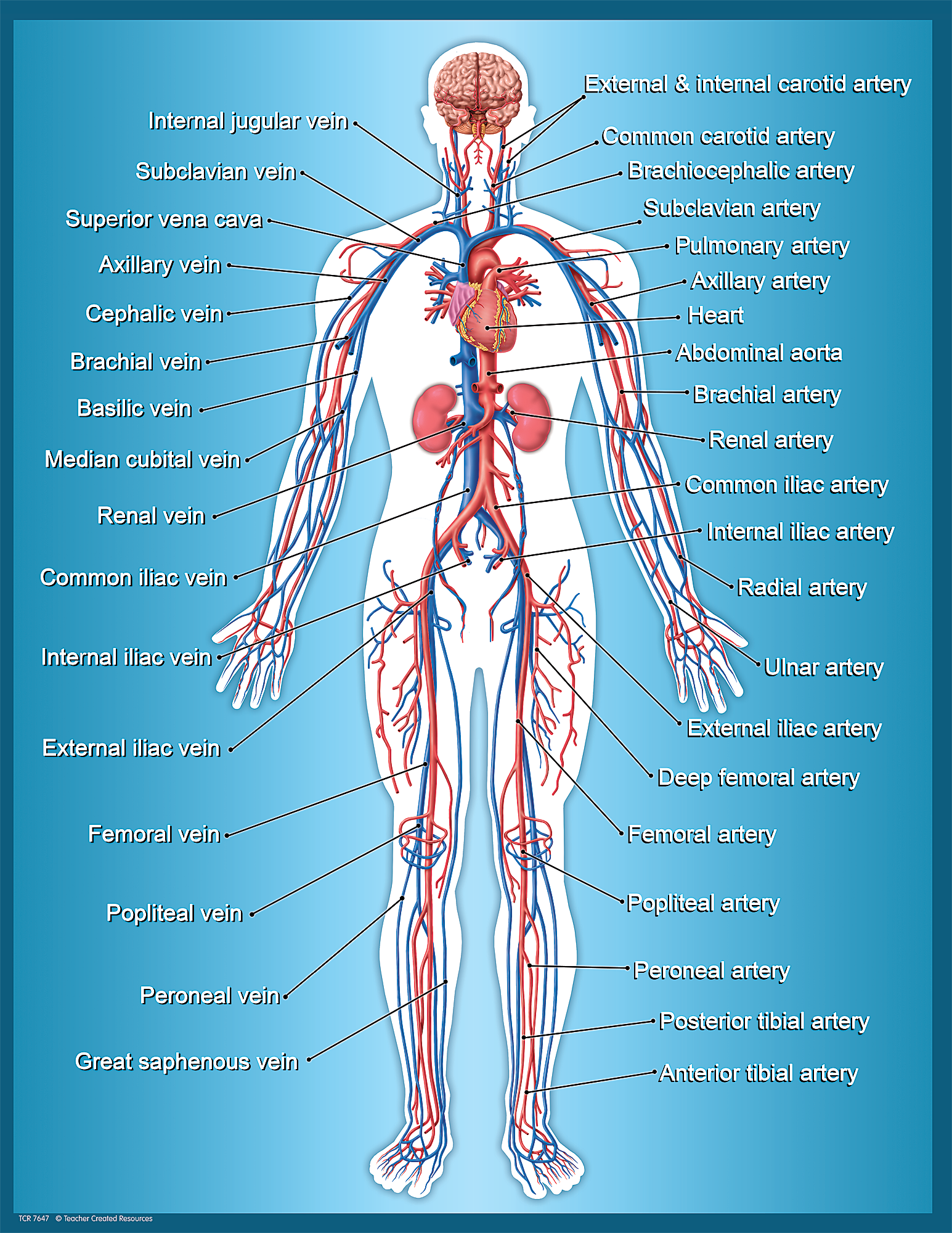
Circulatory System Chart - TCR7647 | Teacher Created Resources
Parts of the Human Brain | Anatomy & Function - Video ... The parts of the brain include the cerebrum, the cerebellum, the brain stem, and the pituitary gland. The brain structure is protected by the skull, which is composed of the cranium and the bones...

01 2012 Human A&P quiz 9 Flashcards - Course Hero
PDF The Human Brain Diagram - Therapist Aid Frontal Lobe • Suppresses socially inappropriate behavior. • Predicts consequences of actions. • Plays a role in the choice between helpful and harmful actions. Temporal Lobe • Assists with the perception and interpretation of sound. • Plays a role in the recognition of objects and visual memory. Occipital Lobe

27 Label The Brain Anatomy Diagram - Wiring Database 2020
(PDF) Pharmacokinetics of Positron-labeled 1,3Bis(2 ... The nitrosoureas are widely used in the chemotherapy of brain tumors, two of the most common being 1,3-bis(2-chloro- ethyl)nitrosourea and 1-(2-chloroethyl)-3-cyclohexyl-1 -nitrosou- rea. ... Pharmacokinetics of Positron-labeled 1,3Bis(2-chloroethyl)nitrosourea in Human Brain Tumors Using Positron Emission Tomography1.

Bald Worm's Blog: Year 5 - Planning: The Fab Four Golden Rules
Human Brain Anatomy - Components of Human Brain with Images Human Brain Anatomy: The brain is composed of the complex network of billions of neurons that are arranged in a specific pattern which is vital to the essential functioning of this organ. Working on the principle of division of labour, different parts of brain are specialized for only specific tasks.
The Anatomy and Physiology of Animals/Nervous System Worksheet/Worksheet Answers - WikiEducator
The Human Brain Atlas at Michigan State University The Human Brain Atlas Keith D. Sudheimer, Brian M. Winn, Garrett M. Kerndt, Jay M. Shoaps, Kristina K. Davis, Archibald J. Fobbs Jr., and John I. Johnson Radiology Department, Communications Technology Laboratory, and College of Human Medicine, Michigan State University; National Museum of Health and Medicine, Armed Forces Institute of Pathology

WMU Psychology Department: Lisa Baker
101 Labeled Brain Images and a Consistent Human Cortical ... Labeling the macroscopic anatomy of the human brain is instrumental in educating biologists and clinicians, visualizing biomedical data, localizing brain data for identification and comparison, and perhaps most importantly, subdividing brain data for analysis.

my red crayon: put your thinking caps on.
The Human Brain - Visible Body The brain gives us self-awareness and the ability to speak and move in the world. Its four major regions make this possible: The cerebrum, with its cerebral cortex, gives us conscious control of our actions. The diencephalon mediates sensations, manages emotions, and commands whole internal systems. The cerebellum adjusts body movements, speech ...

Amazon.com: The Brain Anatomy Poster: Science Lab Anatomy Classroom Supplies… | Brain anatomy ...
Labeled brain anatomy Images, Stock Photos & Vectors Find Labeled brain anatomy stock images in HD and millions of other royalty-free stock photos, illustrations and vectors in the Shutterstock collection.
30 Human Brain With Label - Labels For You

Pin on Neuroscience-brain education
Post a Comment for "45 labels of the human brain"