43 spinal cord model with labels
spinal cord lab model Diagram | Quizlet spinal cord lab model Diagram | Quizlet spinal cord lab model STUDY Learn Write Test PLAY Match + − Created by jasmein_rice Terms in this set (8) anterior horn ... ventral root ... lateral horn ... dorsal root ganglion ... spinal nerve ... posterior horn ... central canal ... dorsal root ... Sets found in the same folder sensory system 79 terms The Brain and Nervous System | Noba It begins as a simple bundle of tissue that forms into a tube and extends along the head-to-tail plane becoming the spinal cord and brain. 25 days into its development, the embryo has a distinct spinal cord, as well as hindbrain, midbrain and forebrain (Stiles & Jernigan, 2010). What, exactly, is this nervous system that is developing and what ...
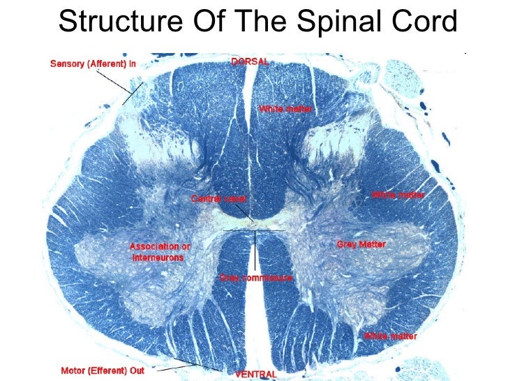
Spinal Cord Segments - Cross-sectional Anatomy - GetBodySmart Start Now. The spinal cord is made up of 31 segments. Each segment gives rise to a pair of spinal nerves. 1. 2. In cross-section (c.s.), the segments appear to be divided into two zones. The outer zone contains many myelinated axons that run up and down the spinal cord. Understand all things spinal cord, white and grey matter with this step-by ...

Spinal cord model with labels
spinal cord anatomy, labeling spinal model Quiz - PurposeGames.com This is an online quiz called spinal cord anatomy, labeling spinal model There is a printable worksheet available for download here so you can take the quiz with pen and paper. Your Skills & Rank Total Points 0 Get started! Today's Rank -- 0 Today 's Points One of us! Game Points 16 You need to get 100% to score the 16 points available Actions Spinal cord: Anatomy, structure, tracts and function | Kenhub The spinal cord is made of gray and white matter just like other parts of the CNS. It shows four surfaces: anterior, posterior, and two lateral. They feature fissures (anterior) and sulci (anterolateral, posterolateral, and posterior). The gray matter is the butterfly-shaped central part of the spinal cord and is comprised of neuronal cell bodies. Anatomy of the spinal cord - e-Anatomy This atlas of human anatomy describes the spinal cord through 18 anatomical diagrams with 270 anatomical structures labeled. It was designed particularly for physiotherapists, osteopaths, rheumatologists, neurosurgeons, orthopedic surgeons and general practitioners, especially for the study and understanding of medullary diseases.
Spinal cord model with labels. MECHANISMS OF HYPNOSIS:: Toward the Development of a Biopsychosocial Model The field lacks an overarching model or framework for organizing the many factors that may contribute to hypnotic responding. We propose in this review that a model hypothesizing roles for biological, psychological, and social factors, and their interactions – that is, a biopsychosocial model of hypnosis—might serve this organizing role. Labeled Spinal Cord Model at Anatomy The ascending and descending tracts of spinal cord transverse section are labelled in detail. Two consecutive rows of nerve roots emerge on each of. It forms a vital link between the brain and the body. Source: The challenge of spinal cord injury (sci) research is to find the right model for testing new treatment strategies. Arm, forearm, and hand: MRI of anatomy - e-Anatomy - IMAIOS Sep 13, 2021 · By moving the mouse cursor over a particular area of the arm or forearm, this area is highlighted and the labels are displayed: anterior, lateral or posterior compartment. On the vertical left menu, a medical illustration of an upper limb skeleton based on a three dimensional (3D) model simplifies the access to the anatomical regions. Spinal Cord - Anatomy, Structure, Function, & Diagram In adults, the spinal cord is usually 40cm long and 2cm wide. It forms a vital link between the brain and the body. The spinal cord is divided into five different parts. Sacral cord Lumbar cord Thoracic cord Cervical cord Coccygeal Several spinal nerves emerge out of each segment of the spinal cord.
9,901 Spinal Cord Stock Photos and Images - 123RF Model of a human spine, spinal columns X-ray C-SPINES : AP, LATERAL showing S/p internal fixation C4, C5 & C6 with plate & screws. There is hypersignal intensity lesion in the spinal cord at C4 to C6 levels, probably myelopathy from compression as described above. Spinal segment with a disk Labeled Brain Model Diagram | Science Trends The medial region of the posterior and anterior lobes function to control fine body movements, taking in input from the spinal cord as well as the auditory and visual systems of the brain. The lateral region of the cerebellum is the largest part of the cerebellum in humans. This region gets inputs from the cerebral cortex. Axis Scientific Spine Model, 34" Life Size Spinal Cord Model with ... This item: Axis Scientific Spine Model, 34" Life Size Spinal Cord Model with Vertebrae, Nerves, Arteries, Lumbar Column, and Male Pelvis, Includes Stand, Detailed Product Manual and Worry Free 3 Year Warranty $76.99 FreeSurfer - Harvard University FreeSurfer software suite. An open source neuroimaging toolkit for processing, analyzing, and visualizing human brain MR images. Visit the Wiki
Nervous System Models - Labeled Brain and Spinal Cord - Pinterest Brain Model Labeled C Carmel Moore Nursing and Medical Neuron Model Brain Nerves C Abby Kate NERVOUS SYSTEM Anatomy Bones Shoulder Anatomy Spinal Cord Model: Dura Mater, Arachnoid Mater, Pia Mater, Ventral Root, Dorsal Root, and Dorsal Root Gangion, Spinal Nerve, Central Canal, Grey & White Matter C Chelsey Renée neurology issues Spinal Cord Diagram with Detailed Illustrations and Clear Labels The spinal cord is one of the most important structures in the human body. In fact, it is the most important structure for any vertebrates. Anatomically, the spinal cord is made up is made up of nervous tissue and is integrated into the spinal column of the backbone. Main Article: Spinal Cord – Anatomy, Structure, Function, and Spinal Cord Nerves GRADE: 5 SUBJECT: NATURAL SCIENCES AND TECHNOLOGY … 9. Learners list any two improvements they will make to their model or to their planning. 10. Depending on the time available you may ask each learner to present their model to the class and to list one improvement they will make. Each learner must hand in the written activities and their model. Use the Formal Assessment Tool and allocate marks as PDF Spinal Cord Classroom Teacher - Duquesne University after reviewing the different parts of the spinal cord, students will draw their own representations of the spinal cord. remind students to include labels on their drawings showing some of the different parts of the spinal cord they learned about! 3. 4. Use spools and thread to make a spinal cord. B o n e M o d u l e t e a C h e r W p a g e S
Spinal cord - Neuroanatomy An Illustrated Colour Text, 4 ed.
Spinal Cord Model - YouTube Dixon discusses Enlargements, cord, pia, dura, mater, denticulate ligaments, 8 spinal nerves, cauda equina, sympathetic chain ganglion , paravetrebral gangli...

Kidney Model – Human Body Help
Spinal Cord in the Spinal Canal (BS 31) · Anatomy models | SOMSO® Spinal Cord in the Spinal Canal Seen from the ventral side, natural size, in SOMSO-PLAST®. The model shows the brain stem and the spinal cord, as well as the nerve branches, up to the coccygeal plexus. On the left side, the sympathetic trunk with its connections to the central nervous system is shown. In one piece. Mounted on a green board.

💠Vertebral column. Spinal Anatomy. 2019-02-13
Sunbeam, Heating Pad Back Wrap with Adjustable Strap … Convenient design: Features 4 heat settings, 2 hour auto off for peace of mind, and a long, convenient 9 foot cord ; Machine washable pad: Super soft, micro mink fabric is machine washable; Simply disconnect the pad from the cord and place in the washing machine
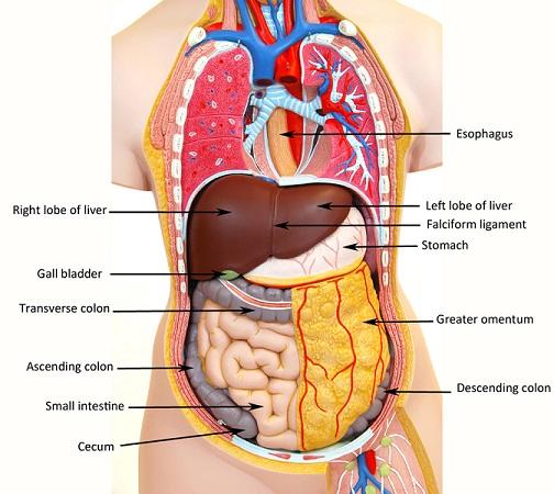
Labeled Human Torso Model Diagram / torso model anatomy labeled 6678046 orig - Top Label Maker ...
Nervous System Models | Spinal Cord with Nerves Models Deluxe Spinal Cord Model (0165-00) Item # DGA65. $369.00 $339.00. Add to cart. Kyoto Kagaku Full-Figure Nervous System Model. Item # KK-A25. Add to cart. Kyoto Kagaku Nerves and Vessels of Arm Model. Item # KK-A144. Add to cart. Physiology of Nerves Series, 5 magnetics - illustrated metal board ...
Print Activity 5: Examining the Human Torso Model flashcards | Easy Notecards
Spinal cord - Wikipedia The spinal cord is a long, thin, tubular structure made up of nervous tissue, which extends from the medulla oblongata in the brainstem to the lumbar region of the vertebral column. It encloses the central canal of the spinal cord, which contains cerebrospinal fluid. The brain and spinal cord together make up the central nervous system (CNS).
Anatomy of spinal cord
Spinal Cord Models - San Diego Mesa College Spinal Cord Models. Click on a photo for a larger view of the model. Click on Label for the labeled model. Back to Nervous System. Spinal Cord (transverse section) Spinal Cord (close up) Spinal Cord (longitudinal view) Label: Label: Label: Spinal Cord (superior ls) Spinal Cord (inferior ls)
Post a Comment for "43 spinal cord model with labels"